Figure 3. Immunohistochemical staining for
CXCR4. Immunoreactivity for CXCR4 was observed in vascular endothelial
cells (A: original magnification 100×) and stromal cells (B:
original
magnification 40×). Double immunohistochemistry for CXCR4
(red) and c-kit (blue). Cells co-expressing CXCR4 and c-kit were
observed in the vascular endothelium (arrows) and in close association
with blood vessels (arrowheads). C: original magnification 100×.
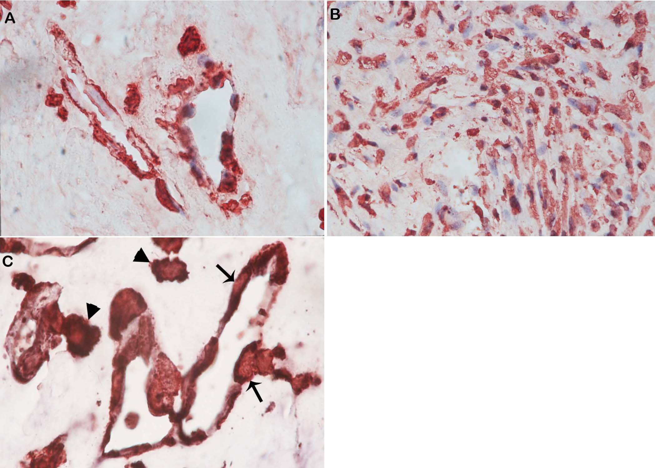
 Figure 3 of Abu El-Asrar, Mol Vis 2010; 16:1098-1107.